







Nuclear Power Summary
25 min read
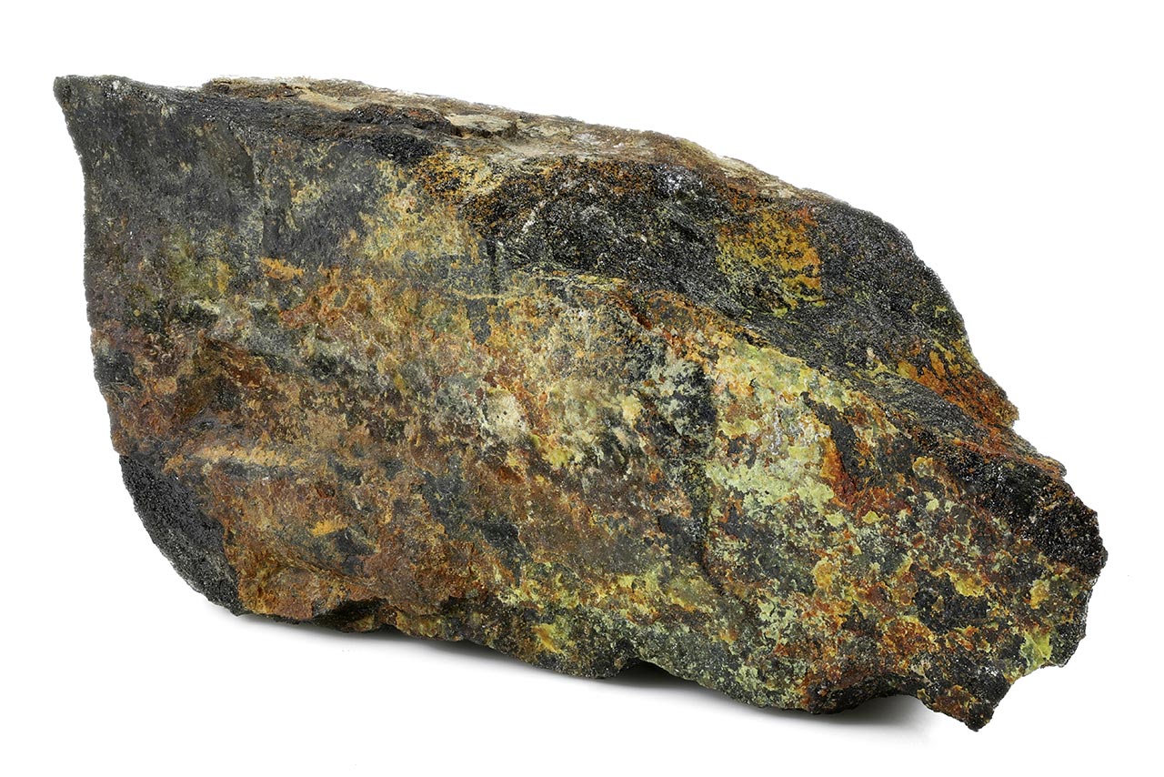
Uraninite, a uranium-rich mineral. Marie and Pierre Curie processed large quantities of uranium ore residues during their research that led to the discovery of polonium and radium.
The era of understanding and subsequently harnessing nuclear energy began in the late 19th century with the discovery of radioactivity and the study of ionising radiation. Numerous experiments with radioactive materials soon led to a deeper understanding of the structure of matter and the composition of atoms.
Radioactivity
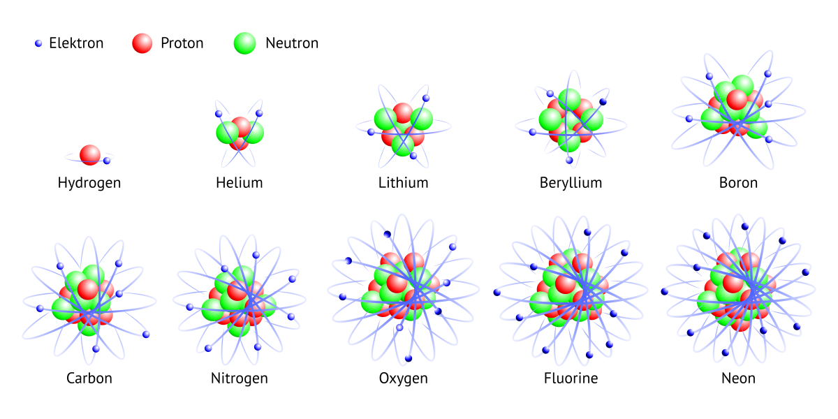
Elements of the first and second periods of the periodic table: hydrogen, helium, lithium, beryllium, boron, carbon, nitrogen, oxygen, fluorine and neon.
Today, we know that radioactivity is a natural component of our environment, present virtually everywhere around us. Its discovery was a pivotal first step — and a fundamental prerequisite — on the path towards the release and utilisation of nuclear energy.
This scientific journey features many remarkable figures. As early as ancient Greece, several centuries BC, Democritus proposed the fundamental idea of matter’s composition, postulating the existence of atoms — small, indivisible, and indestructible particles that constitute the entire universe.
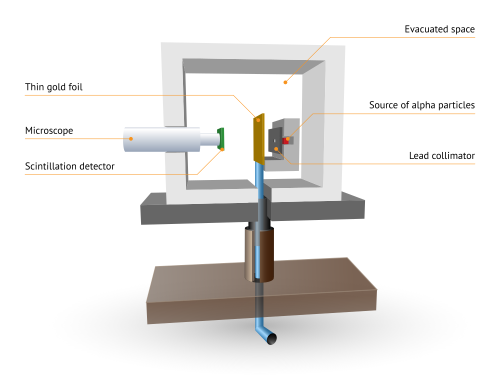
Schematic diagram of Rutherford’s gold foil experiment.
Centuries later, at the end of the 19th century, Wilhelm Conrad Röntgen opened the door to understanding the internal structure of atoms when he studied mysterious cathode ray emissions capable of penetrating solid materials. Shortly thereafter, Henri Becquerel, while experimenting with phosphorescence, discovered that certain minerals — such as uranium salts — could emit invisible radiation without any external energy source. Building on this work, Marie Skłodowska Curie introduced the term “radioactivity” to describe this phenomenon.
Another crucial piece of the atomic puzzle came with J. J. Thomson’s discovery of the electron, a negatively charged subatomic particle. Since atoms were assumed to be electrically neutral, Thomson imagined electrons as raisins embedded in a positively charged “pudding”, a concept later known as the plum pudding model of the atom.
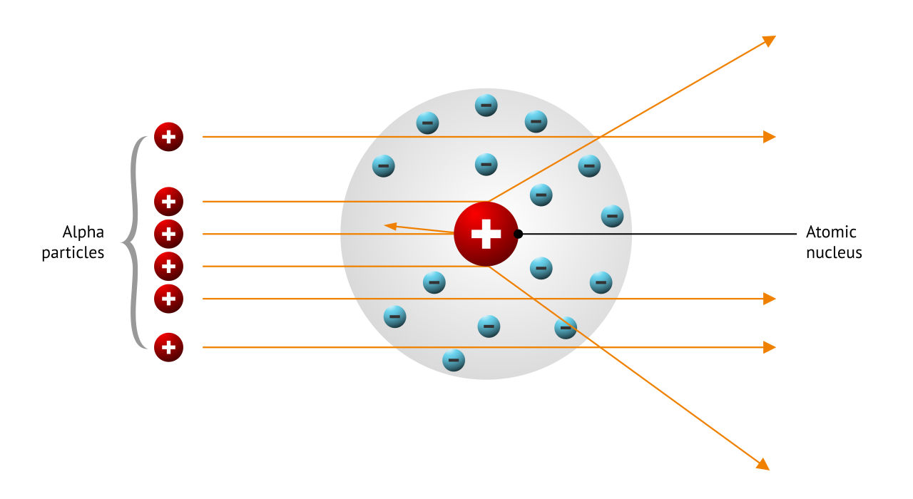
Schematic representation of the scattering of alpha particles by an atomic nucleus.
The nature of radiation was further explored by Ernest Rutherford, who classified it into three types: alpha (α), beta (β), and gamma (γ) radiation. Most importantly, through his famous gold foil experiment, in which alpha particles bombarded a thin sheet of gold, Rutherford demonstrated that nearly all of an atom’s mass is concentrated in a very small nucleus, around which electrons orbit. The nucleus is roughly one hundred thousand times smaller in diameter than the atom itself.
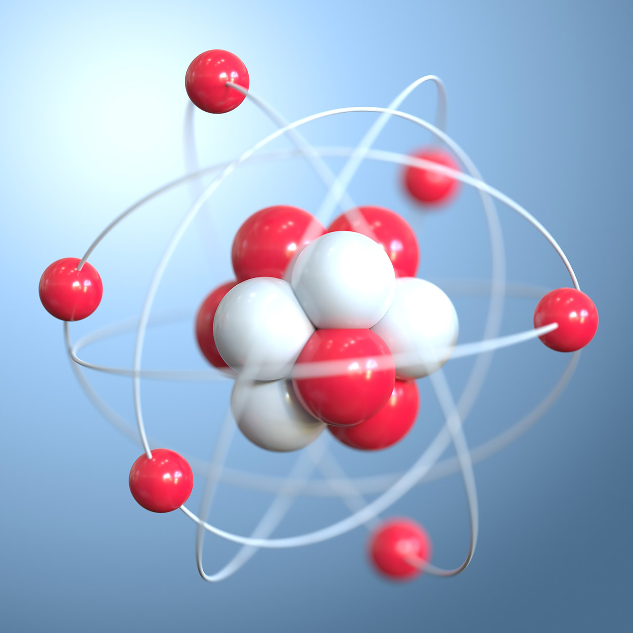
Niels Bohr later refined the atomic model by incorporating early quantum theory, proposing that electrons could occupy only certain permitted orbits with specific energy levels. Although later superseded by quantum mechanics, the Bohr model remains useful for a basic description of atomic structure. To complete the picture, scientists needed to understand the composition of the nucleus itself. The proton, a positively charged subatomic particle, was discovered by Rutherford in the early 20th century, and the final missing piece, the neutron, was identified by James Chadwick in 1932.
You can download the animation of the uranium nucleus model from the Free Downloads section.
The number of protons in an atomic nucleus uniquely identifies a chemical element. Atoms with the same number of protons but different numbers of neutrons in the nucleus are known as isotopes of that element. Isotopes of a given element have very similar chemical properties but differ in mass and nuclear properties.
Some isotopes are unstable, and their nuclei spontaneously transform towards more stable states through a process known as radioactive decay.
{LECTURE_LINKS:2.1:Educational materials on this topic are available in the Learning section. In the chapter “Radioisotopes as Sources of Ionising Radiation”, you can explore video presentations and animated lectures that explain the effects of ionising radiation and introduce many of the pioneering scientists whose discoveries helped reveal the structure of the atom.:Radioisotopes as Sources of Ionising Radiation Learning}
Ionising Radiation
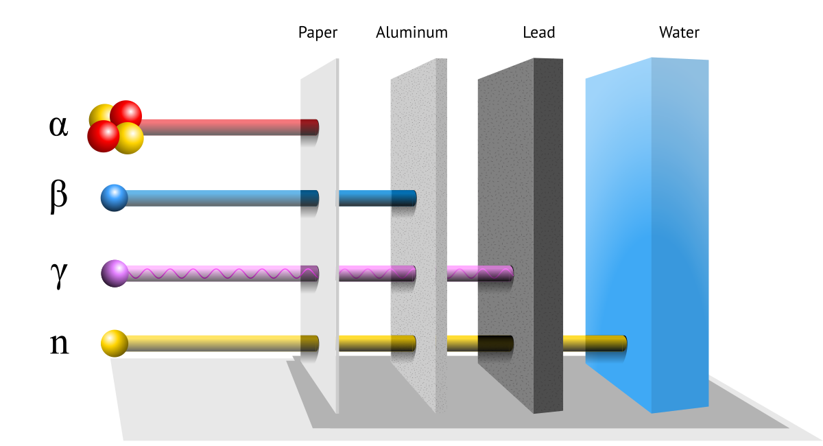
Penetrating ability of different types of ionising radiation.
Free Downloads › Images / Penetrating ability of different types of ionising radiation
Ionising radiation is an inherent and inseparable part of the natural environment. Its primary sources are naturally occurring radioactive elements. The term does not refer exclusively to electromagnetic waves; it also encompasses streams of particles. Ionising radiation has the ability to remove electrons from atoms, thereby producing ionisation.
Alpha radiation (α) consists of a stream of positively charged helium nuclei. It is deflected by a magnetic field and has a very limited penetration depth; even a sheet of paper is sufficient to stop alpha particles.
Beta radiation (β) is essentially a stream of negatively charged electrons or positively charged positrons. Because beta particles are much lighter than alpha particles, they have a greater penetrating ability. A shielding layer of just a few millimetres of aluminium is usually adequate for protection.
Gamma radiation (γ) is also a stream of particles, but in this case it consists of high-energy photons with very high penetration capability. As a form of indirectly ionising radiation, gamma rays are unaffected by magnetic fields and require thick layers of lead to provide effective shielding.
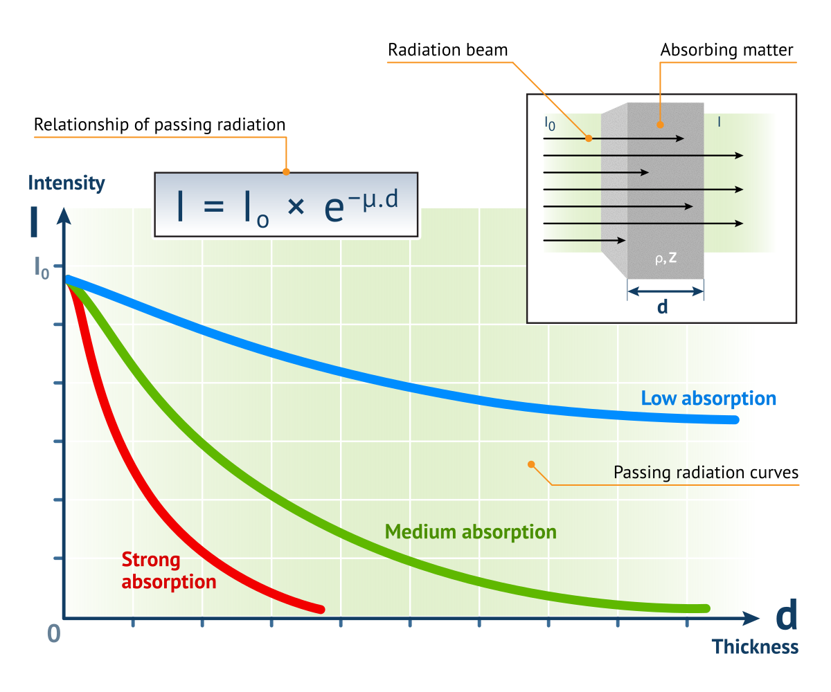
Exponential attenuation of radiation in matter.
Free Downloads › Images / Exponential attenuation of radiation in matter
Other types of ionising radiation include X-rays, neutrons and protons. X-rays and neutrons are indirectly ionising, while protons are directly ionising charged particles. X-radiation is short-wavelength electromagnetic radiation (with a wavelength between 10 nm and 100 pm) that is not generated by nuclear processes, but rather during interactions of high-energy electrons, for example in a cathode ray tube. From the perspective of its origin, X-rays are classified as bremsstrahlung (braking radiation) or characteristic radiation, and they can be shielded using thick layers of concrete or lead.
Neutron radiation is a type of ionising radiation consisting of a stream of free neutrons. It is produced primarily during the fission of heavy nuclei in nuclear reactors or through interactions between alpha-emitting sources and light elements. Like gamma rays, neutron radiation is a form of indirect ionising radiation, as the ionisation is caused by secondary particles generated during neutron interactions with matter.
High-energy protons are a significant component of cosmic radiation that reaches Earth from outer space. Upon colliding with the upper atmosphere, these protons generate a cascade of secondary radiation.
Natural Sources
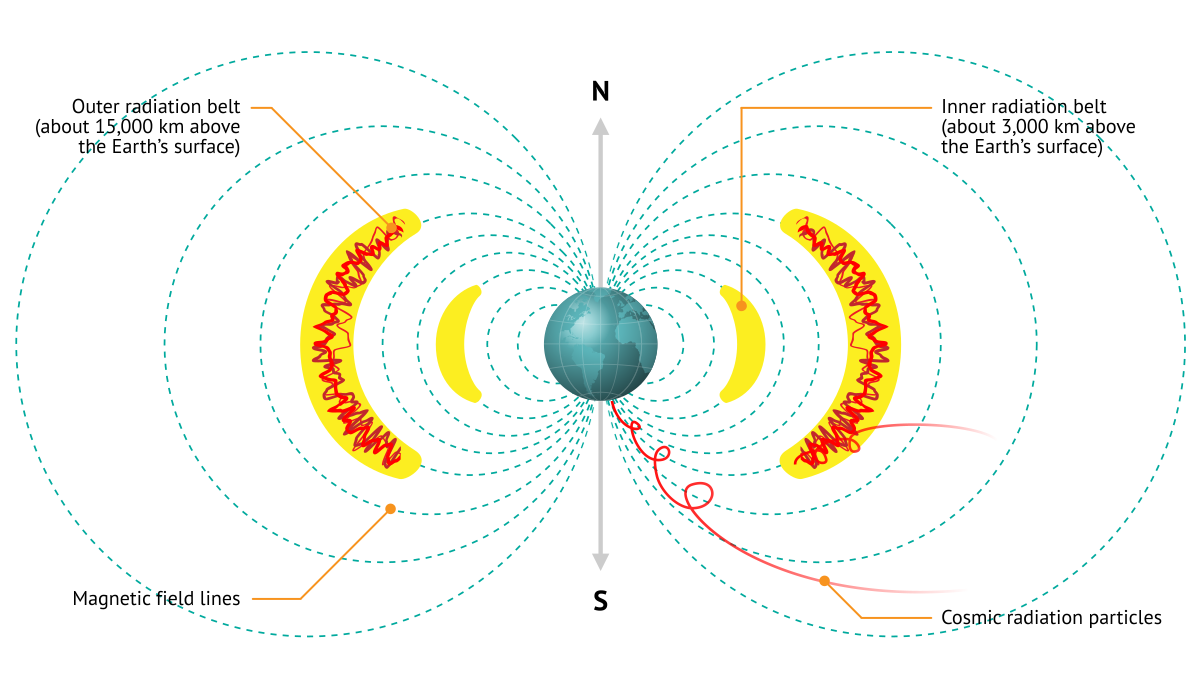
Schematic diagram of the Van Allen radiation belts and the Earth’s magnetic field.
Free Downloads › Images / Schematic diagram of the Van Allen radiation belts
Sources of ionising radiation are present all around us. Radioactive isotopes of various elements occur naturally in the soil, air, water, food, and even within the human body. Collectively, they are referred to as natural sources, and the dose of radiation we receive from them is known as the natural background radiation.
A major part of the total annual dose of ionising radiation originates from natural sources, while the remainder comes from man-made sources, particularly those used in medicine. Exposure from natural sources is far from uniform across the Earth — in some regions, dose rate levels may exceed the global average by one or two orders of magnitude.
One of the fundamental natural sources is cosmic radiation, which reaches the Earth from the Sun or from interstellar space. It consists of a stream of high-energy particles, primarily protons. Only a small fraction of this radiation reaches the Earth’s surface. Most primary particles collide with atoms in the upper atmosphere, generating showers of secondary radiation.
Video: Entry of radon from the ground into residential buildings.
The air and soil are among the most significant natural sources of radiation on Earth. The presence of radioactive noble gases such as radon and thoron, produced by the decay of uranium and thorium, is a major contributor to elevated airborne dose rates.
The occurrence of these gases varies considerably and depends primarily on the geological composition of the underlying strata. The accumulation of hazardous radon inside buildings can be mitigated through adequate ventilation and the installation of radon barriers between the ground and the structure.
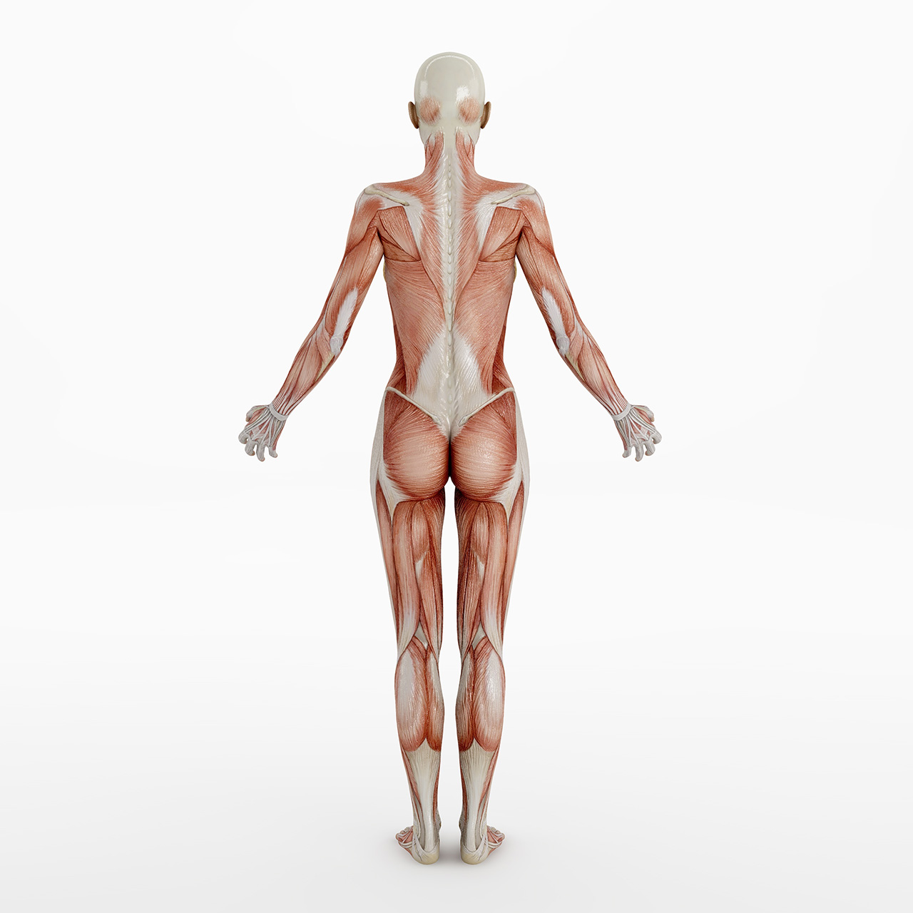
The human body naturally contains small amounts of radioactive isotopes, particularly 40K and 14C.
Rocks and soils contain varying amounts of naturally occurring radionuclides and also contribute to the total radiation dose. Granite, for example, commonly contains higher concentrations of uranium and thorium than many other rock types. Consequently, construction materials produced from such rocks can also serve as sources of ionising radiation. Consequently, construction materials produced from such rocks can also serve as sources of ionising radiation.
Radioactive elements migrate from rocks into the soil, are absorbed by plants, and subsequently enter the entire food chain. As a result, all foodstuffs and drinks we consume exhibit a small degree of natural radioactivity. The specific activity varies — depending on the composition of the soil where plants were grown and on the ability of certain plants or animals to accumulate radioactive elements.
Perhaps unexpectedly, the human body itself is also a source of ionising radiation. It contains radioactive isotopes of elements such as carbon, potassium, and various trace elements, leading to several thousand radioactive decays occurring every second within the human body.
Man-made Sources
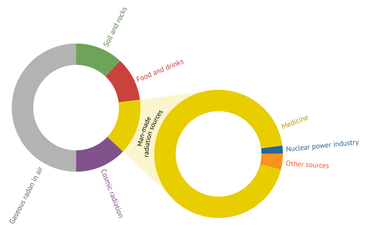
Approximate proportions of the main natural and man-made sources of ionising radiation in the overall dose.
In recent decades, the use of man-made sources of ionising radiation has expanded significantly. These sources are primarily employed in medicine for diagnostic and therapeutic purposes, in industry and energy production, in scientific research, and in many other fields. Exposure from man-made sources is dominated by medical uses of ionising radiation, particularly diagnostic procedures. Relatively small share is mainly the result of radiation protection measures and strict regulatory controls.
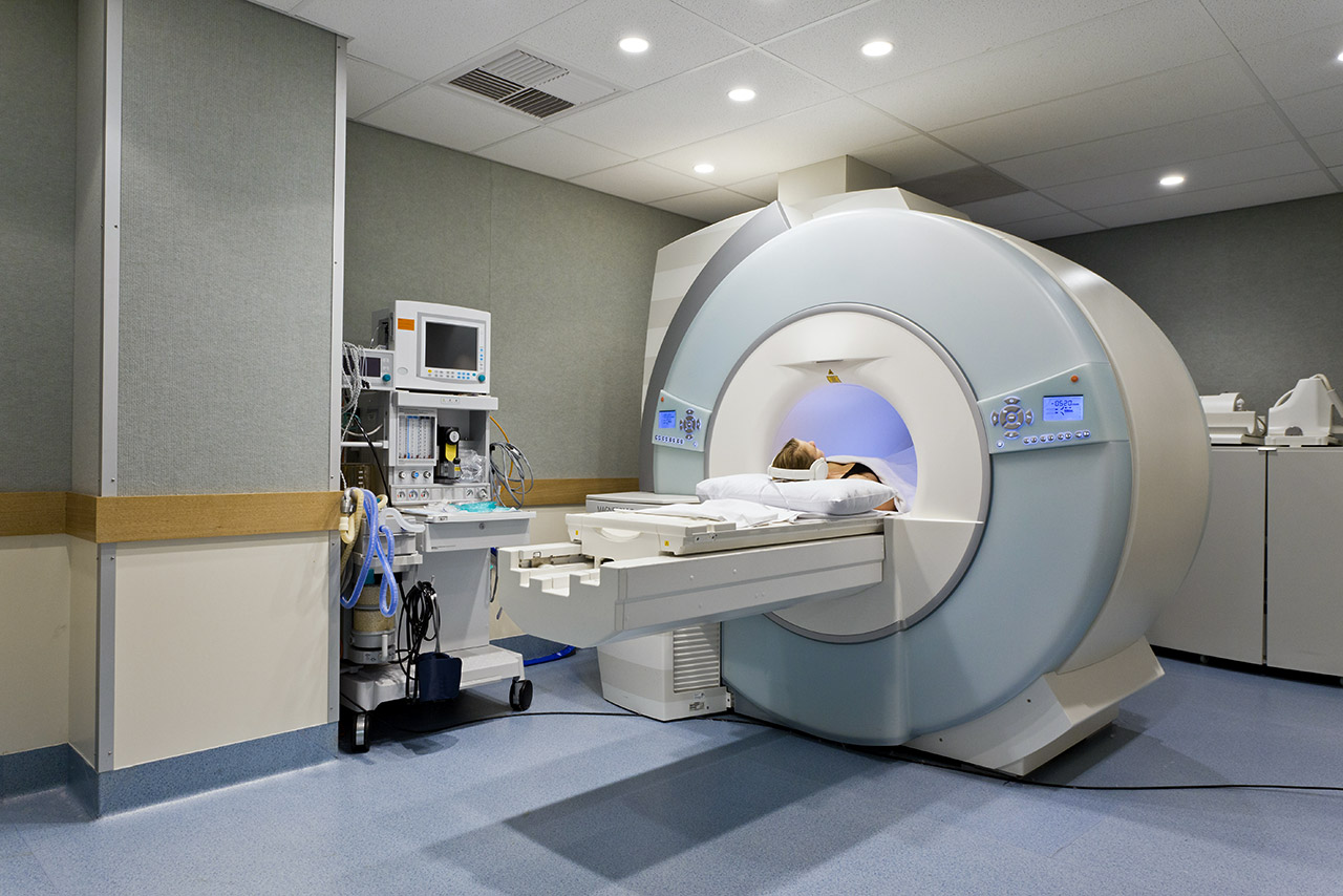
The most prominent area of application for man-made radioactive sources is medicine. Here, radiation is used both for diagnostic purposes — such as radiography, fluoroscopy, and computed tomography (CT) — and for therapeutic treatments, including radiotherapy and brachytherapy.
The administration of liquid open radioactive isotopes into the body, a practice known as nuclear medicine, enables the acquisition of imaging data on the structure and function of specific organs.
Video: Model of a medical X-ray device used for mammography.
High-intensity gamma sources are employed for sterilising surgical instruments, as well as for the protection of cultural heritage objects against wood-boring insects and fungal damage, and even for extending the shelf life of food.
Certain radioisotopes are successfully used as tracers to detect leakages in complex technological systems or to monitor the distribution and absorption of newly developed pharmaceuticals in the human body. Other isotopes assist in the examination of internal material structures, the assessment of weld quality in critical constructions, or the detection of invisible defects in industrial components.
Perhaps the most feared man-made source of ionising radiation is highly radioactive spent nuclear fuel from nuclear power plants. Although it is an intense source of radiation, it is highly concentrated, well shielded from the environment, and stored in strictly regulated interim storage facilities (in countries pursuing direct disposal, intended for eventual disposal in deep geological repositories), resulting in a negligible contribution to public radiation exposure.
Interestingly, coal-fired power plants can also contribute to radiation exposure because coal contains trace amounts of naturally occurring radionuclides, some of which become concentrated in fly ash and other residues during combustion.
Artificial sources of radiation are also encountered in a wide variety of other contexts — from superphosphate fertilisers to uranium-coloured glass. Even domestic smoke detectors utilise trace amounts of radioisotopes as part of their sensing mechanism.
Half-life
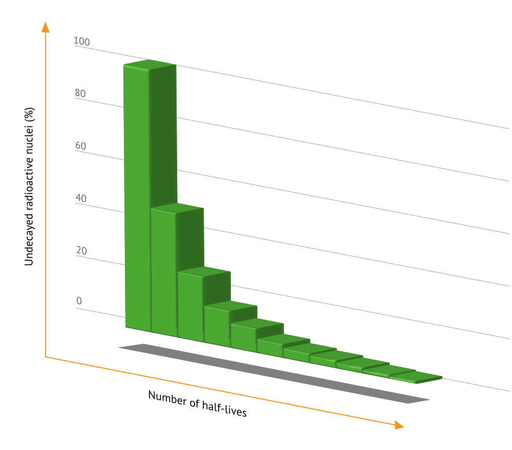
The half-life is the period of time during which approximately half of the atoms of a given radioactive isotope undergo decay, regardless of how many atoms were present initially. This means that half of the original atoms decay during the first half-life, and half of the remaining atoms decay during the second, leaving one quarter of the original quantity. After ten half-lives, about 99.9% of the original radioactive material will have decayed. This fundamental relationship was first described by Ernest Rutherford. The half-lives of different isotopes range from fractions of a second to billions of years.
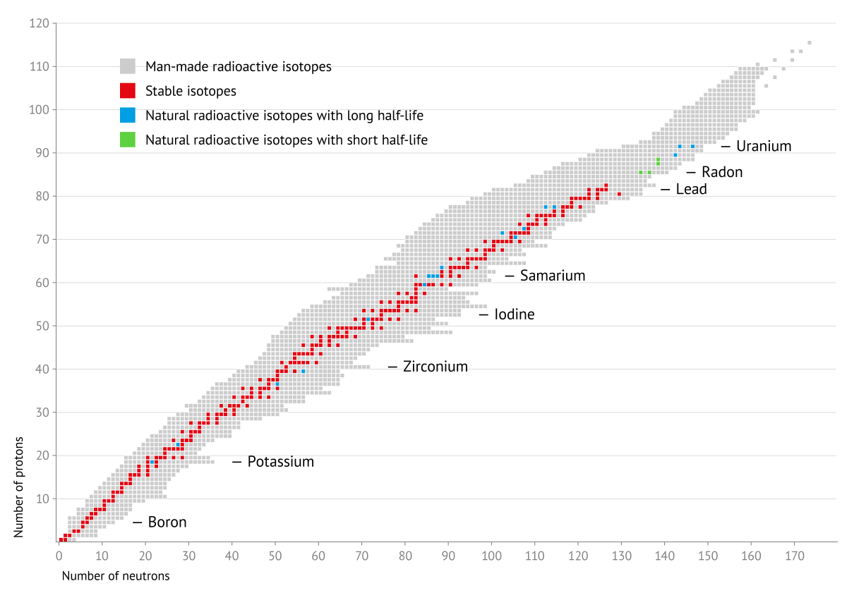
Distribution of stable and radioactive isotopes according to their numbers of protons and neutrons.
In medicine, short-lived isotopes — such as iodine-131 (131I) and technetium-99m (99mTc) — are widely used because they decay rapidly within the patient’s body. The principle of half-life is also the basis of radiocarbon dating, which uses the decay of carbon-14 (14C) to determine the age of biological materials. By measuring the amount of 14C remaining relative to stable carbon, it is possible to estimate how much time has elapsed since an organism died — for example, when it was used to build a ship. While an organism is alive, continuous exchange of carbon with the environment maintains its 14C content in approximate equilibrium with its surroundings. Therefore, if a sample contains half of the original radioactive carbon, its age is one half-life, or 5,730 years.
Much older rocks and minerals can be dated using radionuclides with much longer half-lives. One important method is uranium-lead dating, based on the decay of 238U to 206Pb and 235U to 207Pb. By measuring the proportions of parent uranium isotopes and their daughter lead isotopes in suitable minerals, scientists can determine ages reaching billions of years.
Decay Series
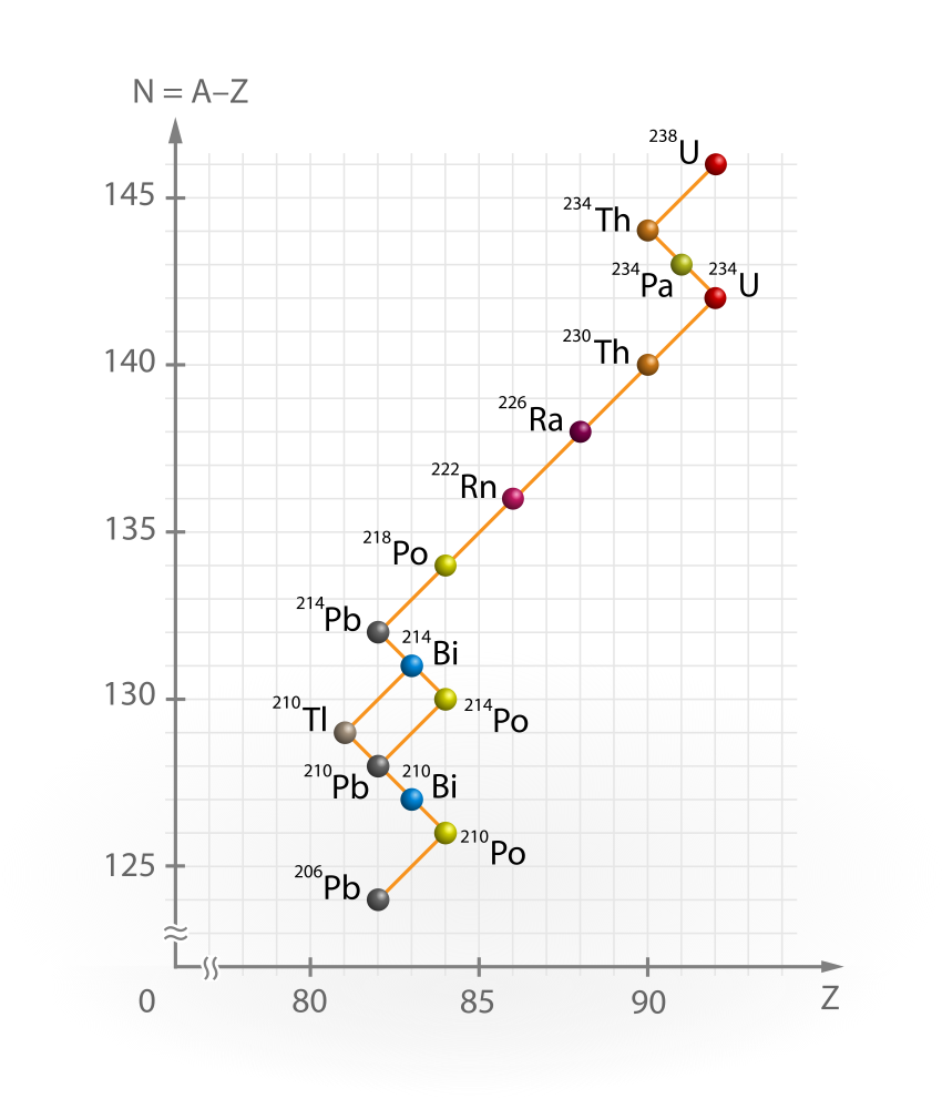
Uranium decay series. The four fundamental radioactive decay series (thorium, neptunium, uranium, and actinium).
Free Downloads › Images / Thorium Neptunium Uranium Actinium decay series
Radioisotopes do not usually decay directly into stable elements. Instead, they transform through a sequence of successive decays that ultimately lead to a stable end product. This sequence is known as a radioactive decay series.
A decay series comprises a range of radionuclides with different half-lives, and it describes their step-by-step transformation. In some cases, a radionuclide in the series can decay via two different pathways, resulting in branching of the series.
There are four fundamental decay series: the thorium, neptunium, uranium, and actinium series. The four series originate from 232Th, 237Np, 238U and 235U, respectively. During the decay process, either alpha particles are emitted — reducing the mass number by 4 — or beta-minus (β⁻) decay occurs, which leaves the mass number unchanged but increases the atomic number by one.
Knowledge of decay series and the relationships between parent and daughter nuclides is important in geochronology and in studies of the origin and history of geological materials.
Quantities and Units
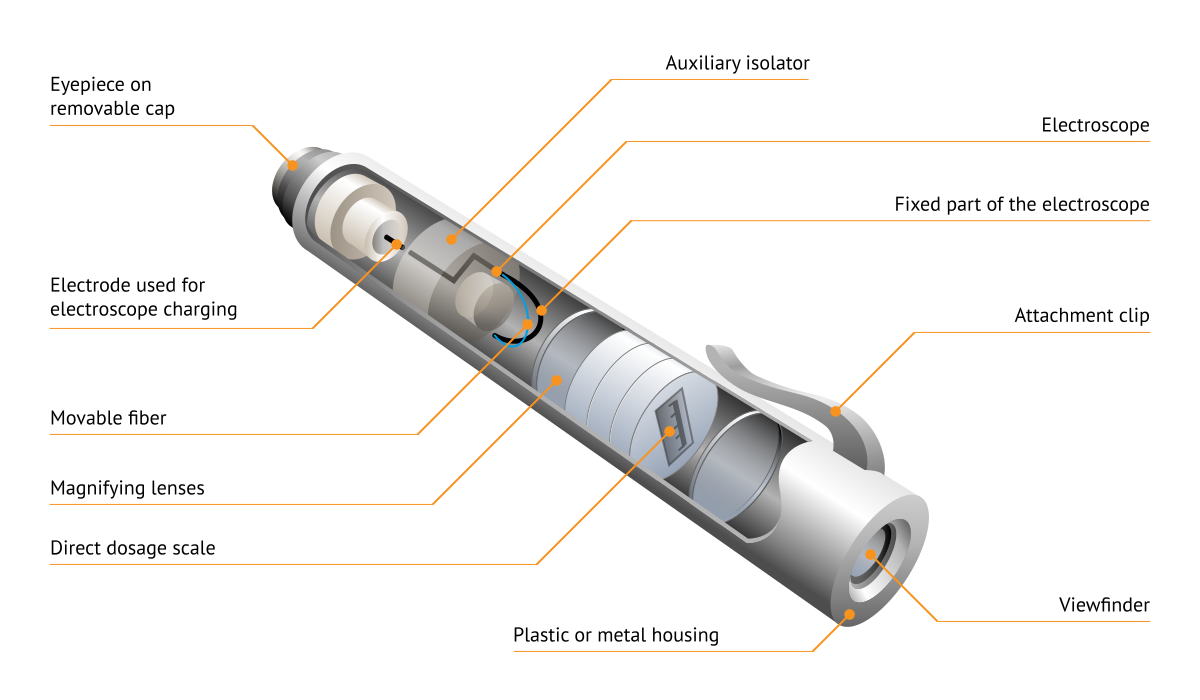
Video: Model of a personal dosimeter.
The behaviour and effects of radioactive substances and ionising radiation are quantified using a set of fundamental physical quantities and measured in corresponding units.
The simple rate of radioactive transformations occurring in a material is known as its activity. The SI unit of activity is the becquerel (Bq), although the older unit curie (Ci) is still sometimes encountered. An activity of one becquerel (1 Bq) corresponds to one radioactive decay per second within a given sample.
1 curie = 3.7 × 10¹⁰ Bq
To evaluate the effect of radiation on a material, the key quantity is the absorbed dose — the amount of energy deposited per unit mass of the substance. It is measured in grays (Gy), where 1 gray (1 Gy) equals 1 joule of energy absorbed by 1 kilogram of material. The older unit of absorbed dose is the rad.
1 rad = 0.01 Gy
Because different types of radiation have different biological effects on living tissue, the concept of equivalent dose is used to represent the biological effectiveness of the radiation. It is measured in sieverts (Sv), or in the older unit rem.
Effective dose also takes into account the different sensitivities of organs and tissues to stochastic effects. Its unit is the sievert (Sv), and it is widely used in radiation protection to compare overall exposures.
The dose rate expresses the amount of radiation dose received over a specific period of time, such as per minute or per year. When assessing radiation exposure in humans, the equivalent dose rate is expressed in Sv/s. Because the sievert is a relatively large unit, microsieverts per hour (µSv/h) or millisieverts per year (mSv/year) are more commonly used.
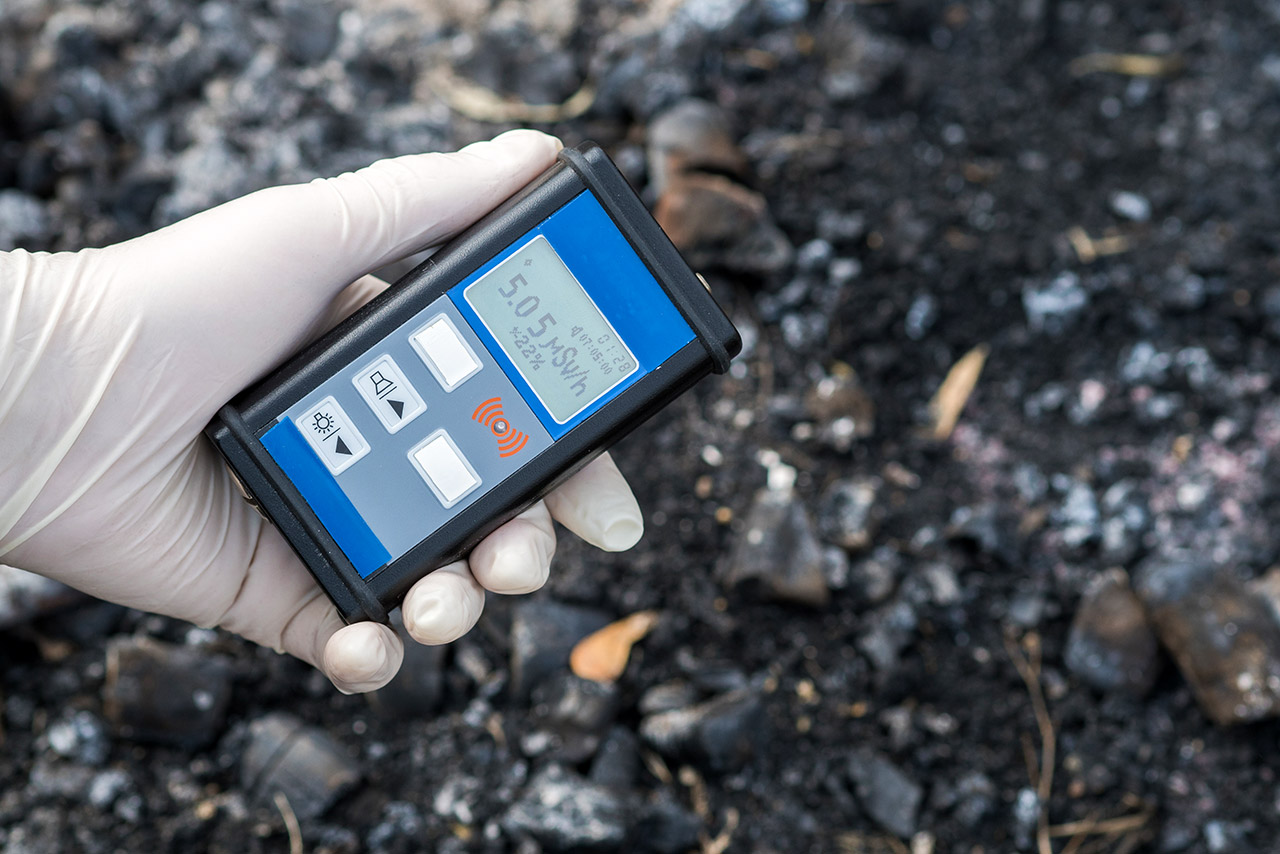
Various types of detectors are used to detect ionising radiation in both laboratory and operational environments. Despite differences in design, all detectors operate on the same basic principle: radiation passing through the detector produces a measurable physical effect. The most common types include: Scintillation detectors; Track detectors; Semiconductor detectors.
Other detectors, such as ionisation chambers, measure the electrical charge produced when radiation ionises a gas. A Wilson cloud chamber operates on a different principle, making the tracks of charged particles visible through condensation in a supersaturated vapour.
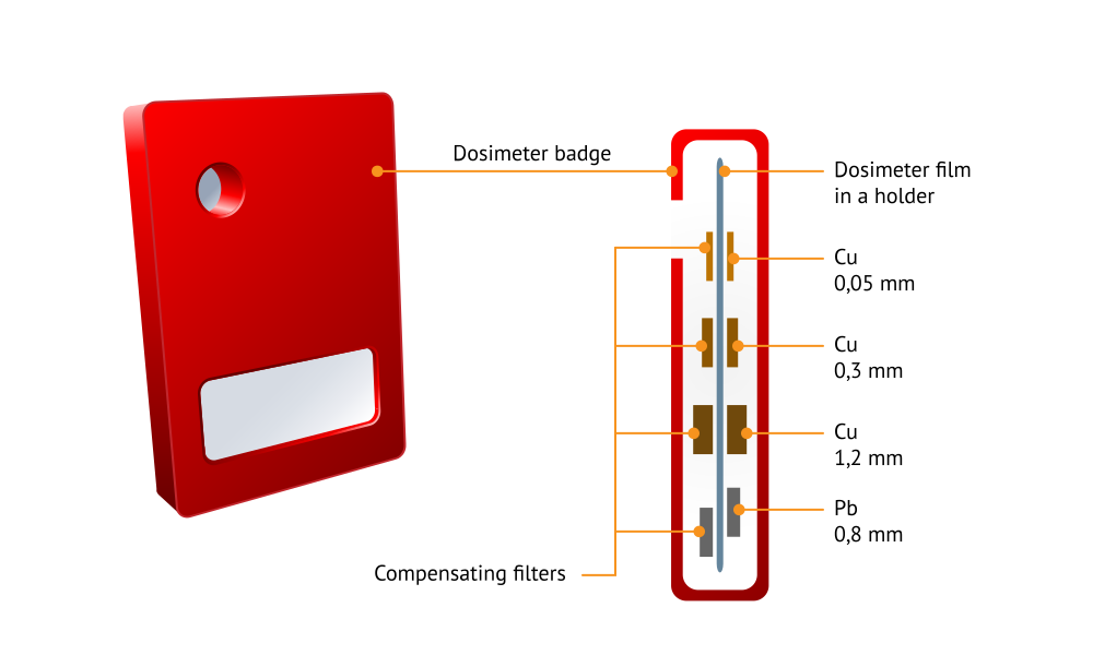
To measure the radiation dose received by a person, dosimeters are employed.
In a film dosimeter, particles passing through the device cause gradual darkening of the photographic film — the greater the radiation dose, the more intense the darkening.
A thermoluminescent dosimeter (TLD) stores part of the energy deposited by radiation in trapped electronic states. When the material is subsequently heated, the electrons are released and light is emitted, with its intensity related to the absorbed dose.
Effects of Ionising Radiation
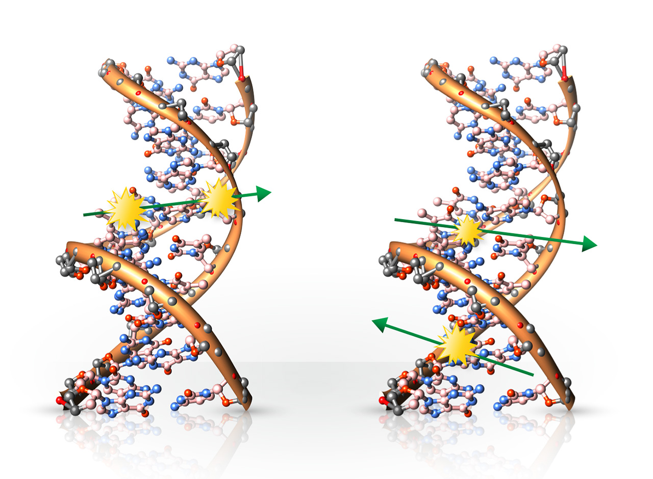
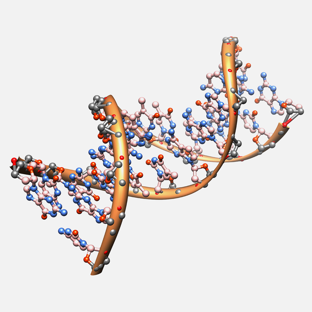
DNA damage caused by ionising radiation — one or both strands of the DNA double helix may be broken.
Ionising radiation has the ability to dislodge electrons from atoms through which it passes. This interaction can lead to molecular damage or structural changes in matter. In the case of living cells, such radiation can severely damage or even destroy the cell. Cells can repair many types of radiation-induced damage. Incorrectly repaired or unrepaired DNA damage in surviving cells, however, may result in mutations and can potentially contribute to the development of cancer.
Some studies have suggested that low doses of radiation might stimulate protective biological responses, an effect known as radiation hormesis. However, this hypothesis remains scientifically controversial and is not used as a basis for radiation protection standards. Conversely, high doses of radiation can cause severe tissue damage, organ failure, and ultimately death.
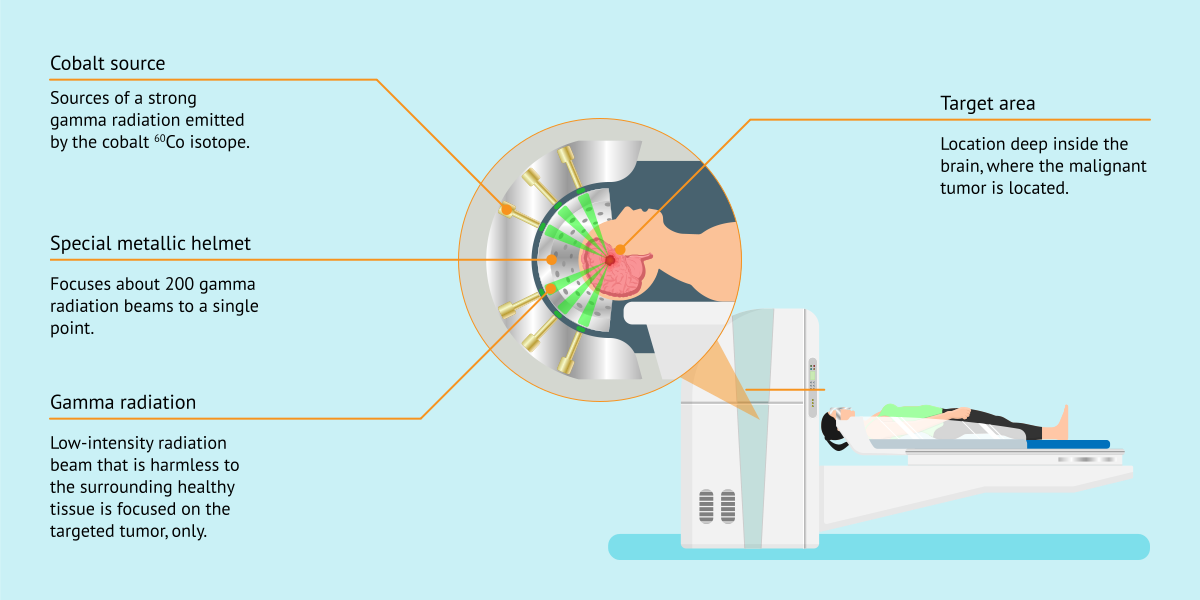
Principle of the Gamma Knife for targeted treatment of lesions in the brain.
Free Downloads › Images / Gamma knife Cobalt-60 radiotherapy unit
In medicine, high radiation doses are deliberately delivered to tumours in radiotherapy. Treatment is carefully planned to destroy cancer cells while limiting the dose to surrounding healthy tissues. Radiation may be delivered externally or by placing radioactive sources in or near the tumour. Highly focused beams can also be directed at a target from multiple angles, as in the Gamma Knife.
Low-dose exposure to radon is also used therapeutically in some spas, particularly in the treatment of rheumatic and musculoskeletal disorders.
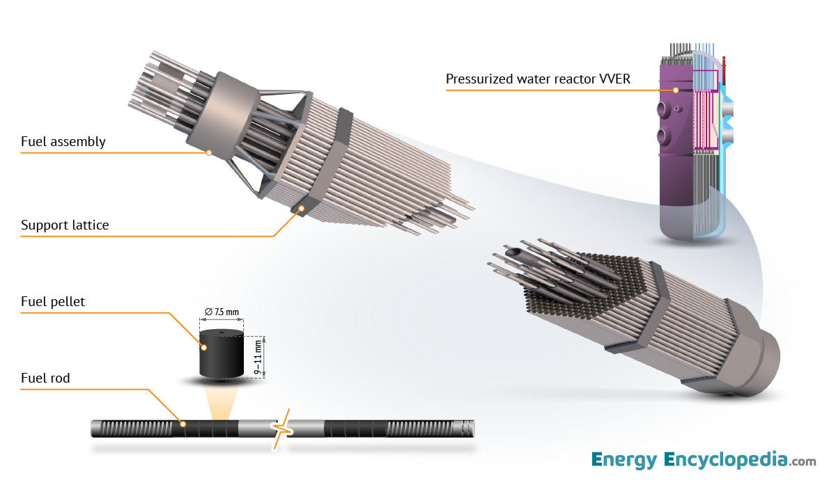
If you are interested in the design of the pressure vessel of the most widely used PWR reactor, you can view it online in the 3D Models section or download the embed code from Free Downloads.
Online 3D Model › NPP PWR / Reactor
Free Downloads › Online 3D › NPP PWR / Reactor
Irradiation has specific applications in the modification of industrial materials. Radiation-based technologies enable the production of materials with entirely new properties without the use of undesirable chemical additives. Radiation processing can be used to initiate polymerisation, cross-link polymers, produce polymer foams,cure coatings, alter the properties of textiles, or change the colour of glass. Radiation cross-linking of polyethylene is also used in the manufacture of heat-shrinkable products.
On the other hand, fast neutrons can gradually embrittle the steel of a reactor pressure vessel. Reactor vessels are therefore made from carefully selected steels, and changes in their mechanical properties are monitored throughout the operating life of the plant.
Radiation Doses and Activities
Our bodies are exposed to a certain level of radiation dose during almost every activity. For example, consuming a single banana exposes the body to a dose of approximately 0.1 µSv. By contrast, living for one year in the Brazilian city of Guarapari would result in exposure to a dose of around 100 mSv.
There is also a significant variation in the activity of various products and materials. For instance, 1 kg of coffee has a radioactivity of about 1,000 Bq, while americium contained in a household smoke detector reaches approximately 30,000 Bq. By comparison, 1 kg of natural uranium has an activity of approximately 26 million Bq.


Robie, your AI assistant